


There are various electromagnetic waves, some of which are visible and others that are not. Radio waves, UV rays, and microwaves are the most familiar ones. X-rays are made possible by these invisible waves of electromagnetic energy, and if you want to take an x-ray, you will find the best x-ray centre in Kamothe if you search “x-ray lab near me“. But first, let’s take a look at what x-rays are and the mechanism behind it.
X-ray imaging is used to obtain images of the inside of your body because different tissues absorb different levels of radiation, the images show different parts of your body in distinct shades of black and white. Bones appear white because calcium absorbs the most x-rays. Fat and other soft tissues absorb less and thus appear gray in color and the lungs seem black because air absorbs the least.
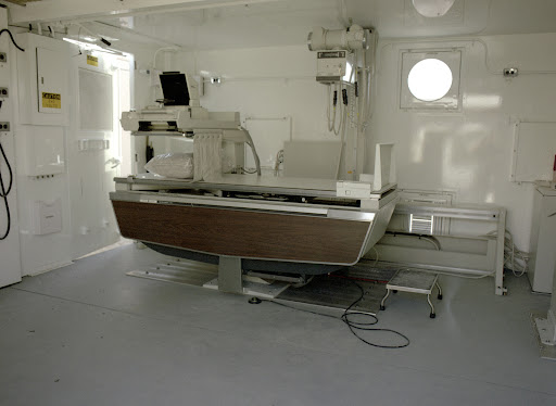
X-rays are mostly employed in medicine and dentistry, and here’s the best diagnostic centre in new Panvel where you can visit to diagnose fractured bones and fractures, ingested objects, arthritis-related bone deterioration, and lung infections. CT scanners, or computed tomography, also employ X-rays. This modality employs numerous X-rays to produce a layer-by-layer picture during a single scan.
The X-ray machine, commonly known as an X-ray tube, generates X-rays, and there is no external radioactive material involved. Radiographers can modify the parameters of the X-ray beam produced by changing the current and voltage settings on the X-ray equipment, and different X-ray beam spectra are used on various bodily areas.
A slight rise in filament voltage causes a massive increase in tube current, accelerating high-speed electrons from the hot filament negative cathode within a vacuum towards a positive tungsten target electrode. This anode rotates to dissipate the heat produced. X-rays are produced within the tungsten anode, and an X-ray beam is directed at the patient.
When a high energy electron collides with an inner shell electron, they are both ejected from the tungsten atom, leaving a “hole” in the inner layer. This is filled by an outer shell electron, with an X-ray photon emitted as energy loss.
A spectrum of X-ray energy is formed within the X-ray beam due to characteristic and bremsstrahlung radiation creation.
This spectrum can be altered by adjusting the X-ray tube’s current or voltage settings or filters to filter out low-energy X-rays. Radiographers can use this technique to apply distinct spectra of X-ray beams to different bodily areas.
Find an expert diagnostic centre in New Panvel to get an x-ray done. Simira Healthcare offers the most significant quality services of x-ray and other diagnostics. Visit Simira Healthcare, the best and most reliable x-ray centre in Kamothe.
Copyright © Simira Healthcare Private Limited 2024. All Rights Reserved


This will close in 0 seconds
This will close in 0 seconds


Leave a Reply